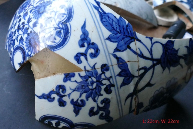
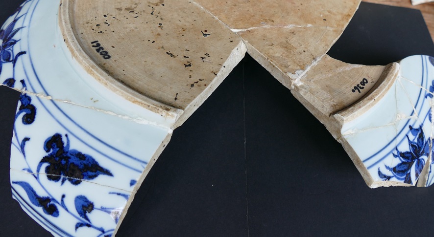
We have investigated two shard samples from Yuan period. One is from a Meiping vase. The shard is with length 22cm, width 22cm, and thickness 1cm.
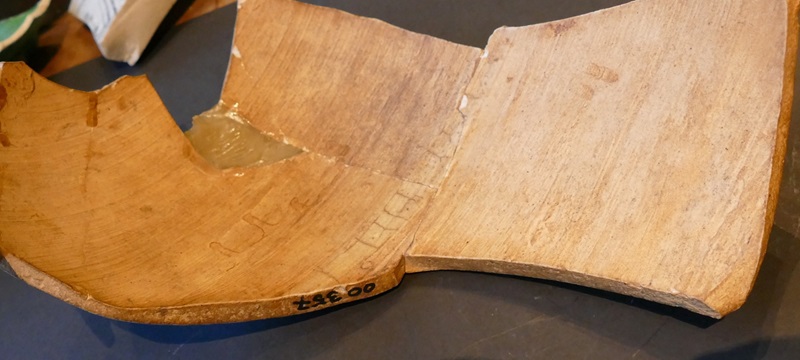
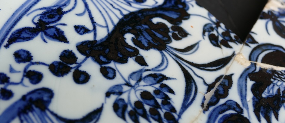
The other one is from a big plate with pond motif, which is with length 38cm, width 22cm, and thickness 1cm.
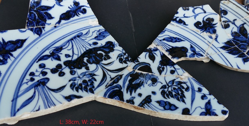
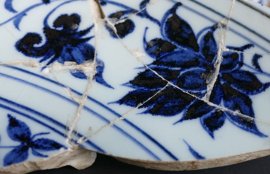
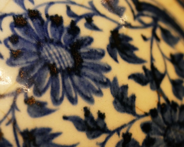
With naked eyes, the iron spot from these two samples showed differently. Generally, the iron spot from the latter shard 019 appeared more obvious and particularly, many blue dots on the blue areas attracted eye sights.
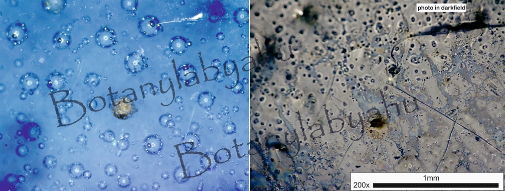
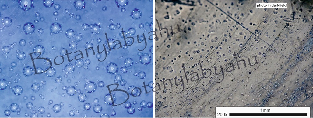
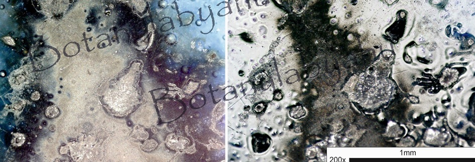
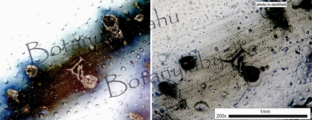
1 Black specks / iron spots from Meiping vase
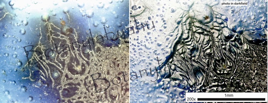
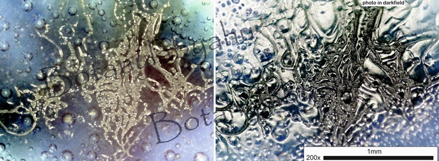
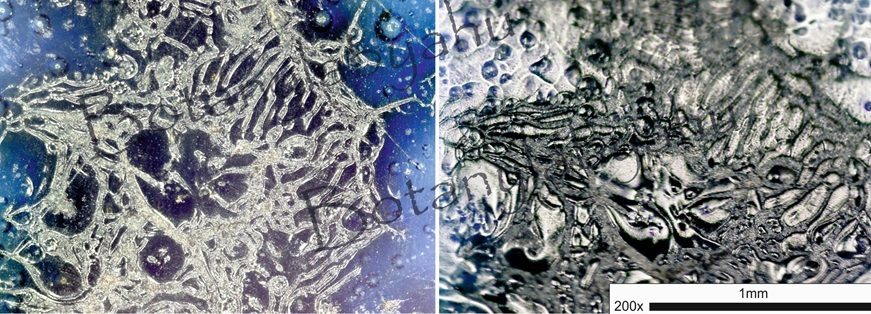
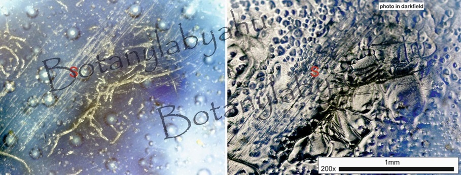
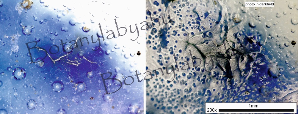
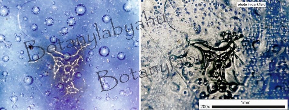
Most of the iron spots showed net-like structure with various sizes. Under the darkfield microscope the net-like structure appears black, which is darker than the silver color of glaze area around. Possibly there accumulating CoFe2O4 along these net-like structure. Through the net-like structure the glossy glaze can be seen, which shows silver under the darkfield microscope. Meanwhile, some traces of scratch (marked as ‘S’ on the picture) on the surface can be seen.
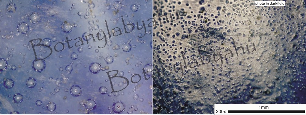
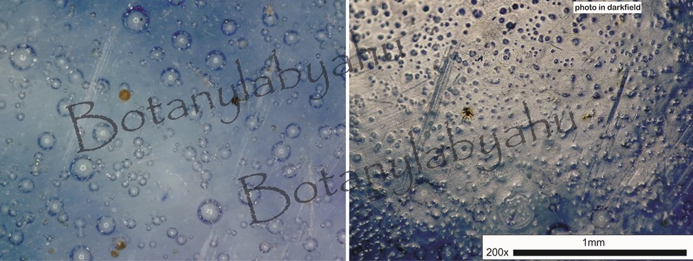
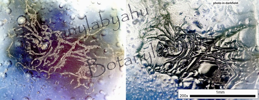
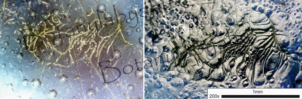
2 Black specks / iron spots from big plate
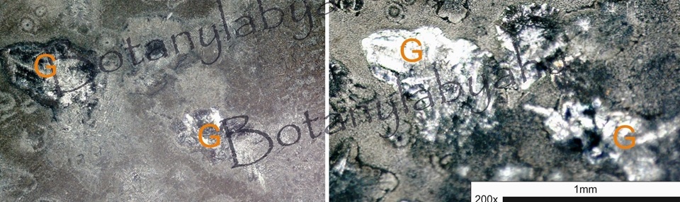
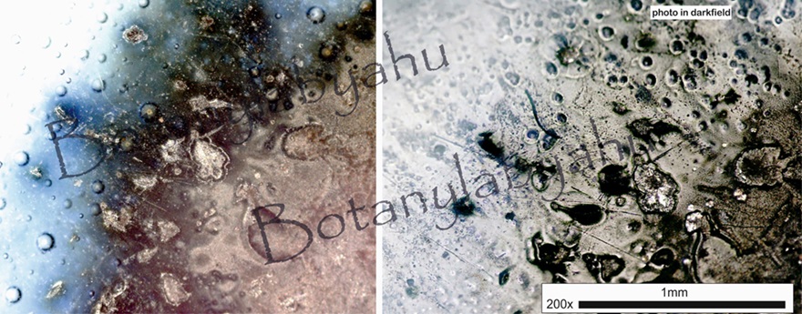

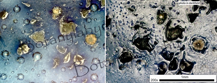
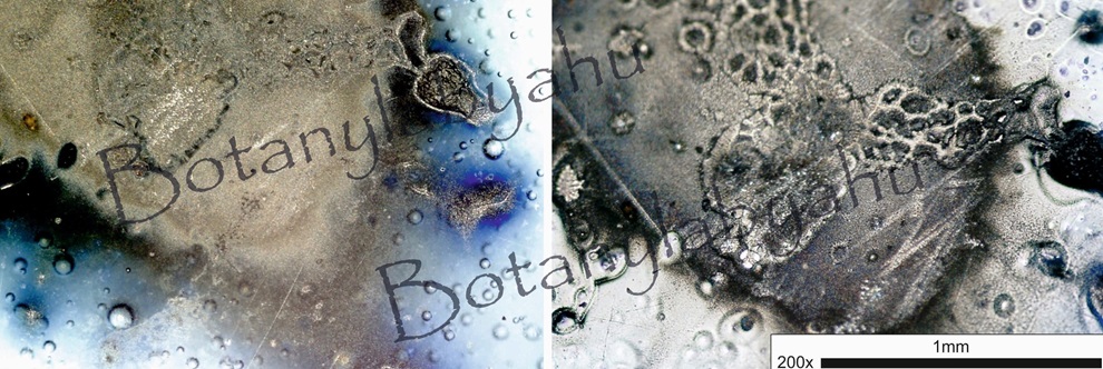
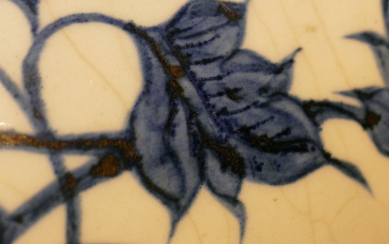
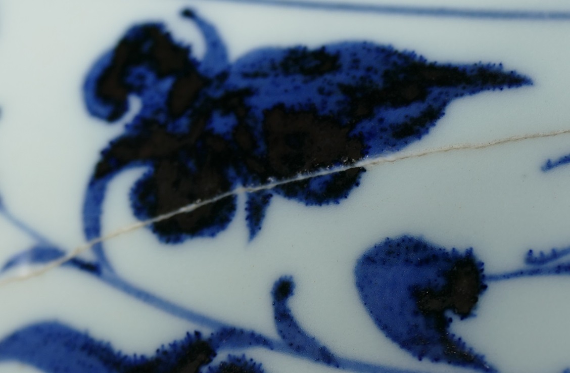
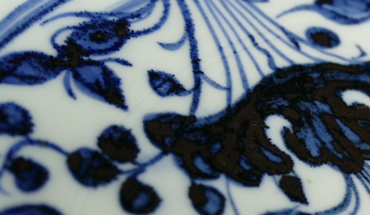
Only a few of net-like structures can be seen. Most of the iron spots show rust-like structure. On the big flaky iron spots some small areas show glaze (silver color under the darkfield microscope). With our naked eyes we can see that lots of blue dots spreading on the blue areas. Under the microscope we can see that these blue dots are from some grain-like material. These grains show various sizes. Moreover, these grains also appear on the flaky spots. So far there is no study mentioned about this phenomenon. According to our investigation, the primary idea about these dots we could get is: during preparation of cobalt pigment, some material containing relatively high Si2+ was added extra in. Moreover, this material was not integrated well into the glaze. On photos ‘G’ presents glossy glaze on the surface.
3 glaze on the surface